Most of us will have spent some of our science lessons at school peering down a microscope, looking perhaps at dividing cells, leaves or sections of organs.
The moment when you look through the eyepiece can be like entering another world, one in which everything operates on a totally different scale.
That different sense of scale is possible thanks to the miscroscope’s most important part: the lens. This precisely curved piece of glass is what takes the light coming off the specimen, however tiny, and focuses it on the eyepiece where you can see it at a scale that makes sense.
No lens, no microscope. Or so you’d think.
But a professor in Dubai is hoping to change that, with a new form of microscopy that does away with the lens.
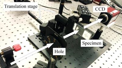
Dr Suhas Veetil, an assistant professor at Amity University in Dubai, is working on Fourier Ptychographic Microscopy (FPM), a new method that some researchers view as a major breakthrough in the field of microscopy.
Rather than using a lens to focus the light from the specimen, it looks at how the specimen diffracts – or scatters – light shone through it on to a recording plane behind, before complex mathematical processing is used to create an image of the specimen.
The method is not just restricted to looking at light, either: it can also detect how electrons, X-rays or lasers are scattered by a specimen.
To understand how the process works, Dr Veetil says people can imagine it as being like throwing a handful of sand at an object, and seeing what pattern emerges.
“There’s an object. I’m throwing some sand through this object. It hits the object and it’s scattered to the ground,” he says.
“Looking at this pattern of the sand on the floor, you can think about the particular object the sand hit. This is what the diffraction pattern tells you. From the footprint you can go back to [work out] what the object was. This is called phase retrieval.”
In simple terms, this process of phase retrieval involves taking the basic, blurred image produced by the object – which in no way resembles the object itself – and processing it by using complex mathematical formulae called algorithms several times.
The scientist initially creates a rough guess of what the image looks like, and uses this as a reference point when processing the image. The processing continues until the picture that emerges is the same as the image that went in.
But why do away with the lens in the first place? According to Dr Veetil, who hails from India and has worked as a researcher in Germany and South Korea, standard microscopes have a number of drawbacks.
A key problem with lenses, he says, is that there is a trade-off between the level of detail that can be seen through the lens – the resolution of the image produced – and the amount of the specimen that can be viewed all at once.
Aberrations in the lens can require the use of expensive optical equipment, while traditional electron microscopes – which look at the scattering of electrons, instead of light, by a specimen – are held back by power-supply instabilities that cause variations in the energy of electrons, he says. These can affect the quality of the image produced.
Also, the staining that is often needed to highlight features of an object that is being viewed with a lens can be damaging to the specimen, particularly biological material such as cells. With FPM, no staining is needed.
Dr Veetil says the method could represent one of the biggest steps forward in microscopy since the introduction of the electron microscope in the 1920s and 1930s, and the creation of atomic force microscopes, which use tiny probes to map the surface of objects, in the late 1980s.
“This is going to be another breakthrough in the field of microscopy. That’s the potential it has,” he says.
A whole specimen can be scanned, in great detail, at once, removing the need to take several pictures to create a detailed record of the whole object, something that can take several tens of minutes.
And the potential uses of FPM are many. Scientists can examine anything from biological specimens such as cells to crystalline structures – such as semiconductor wafers – to check for defects. Almost any nanostructure that scatters X-rays, electrons, lasers or normal light can be assessed.
The focus of Dr Veetil’s work, which is conducted in collaboration with scientists at Shanghai Institute of Optics, is in refining the mathematical steps used to process the image. When developing the algorithms, the key issues are the need to improve the contrast in the image, and increasing what is known as the signal-to-noise ratio, which means getting more useful information from the specimen without collecting additional unhelpful information that the microscope generates.
There are now companies trying to commercialise the techniques of FPM, although Dr Veetil cautions that it will probably be a while before they can be used routinely.
“People may have to wait for some time, until this is ready on a table-top machine where this screening is done automatically and you get the image,” he says.
Ultimately, FPM is likely to be of greatest use in research centres, rather than in facilities that screen large numbers of samples.
“You may not need this in normal hospital labs. They are not going into that much detail. [It is] more where such molecules are developed – pharmaceutical laboratories, like research-and-development institutions where research is taking place,” he says.
“Where people need to see the structure, where they need to change the structure, to manipulate the chemical behaviour, the physical behaviour.”
It is in research laboratories of the kind where ptychographic microscopy will probably prove most useful that many of tomorrow’s scientific breakthroughs are likely to be made.
So although ptychographic microscopy is highly specialised, it could prove useful in helping science to progress, thanks in part to researchers such as Dr Veetil.
newsdesk@thenational.ae


